FIRST CLICK HERE AND READ OUR OVERVIEW

Originally published in February 2004 icon
Melanoma of the eye (ocular melanoma) is a rare cancer. There are no accurate figures on how many people are affected by it a year in the UK. Melanoma is a cancer that develops from cells called melanocytes.
These cells produce melanin, the dark-coloured pigment which is responsible for the colour of our skin.
Melanocytes are found in many places in our body including the eye.
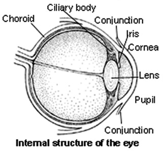
Ocular melanoma can develop in one of several places within the eye. The most common is along the uveal tract of the eye (uveal melanoma), which includes the choroid, ciliary body and iris. The choroid is part of the lining of the eyeball.
The ciliary body extends from the cboroid and focuses the eye by changing the shape of the lens. The iris is the coloured disc at the front of the eye, which controls the amount of light entering the eye. All these structures are heavily coloured with melanin. Melanoma can also occur in the thin lining over the white part of the eye (the conjunctiva) or on the eyelid, but this is very rare.
The cause of this kind of tumour is unknown. However, we do know that exposure to UV rays (either from the sun or sunbeds) increases the risk of developing melanoma of the skin. People whose skin burns easily (those with fair or red hair and blue eyes) are most at risk. However, it is not yet known whether there is any link between UV ray exposure and the development of melanoma of the eye.
Signs and Symptoms
These include blurred vision, flashing lights, shadows and misting of the lens of the eye (cataract). Often no symptoms are noticed until the tumour is quite large. All of these symptoms can be a sign of other eye conditions, but normally an eye specialist can diagnose these tumours quite simply and painlessly.
Treatment
A number of different treatments can be used, depending on the size, cell type and position of the tumour - as well as the general health and age of the patient, and the level of vision in both eyes. The aim is to destroy the cancer cells, stop the cancer coming back and save as much vision as possible.

Recent developments in radiotherapy have made it possible to completely or partially preserve the patient’s vision

Radiotherapy
High-energy rays are used to destroy the cancer cells. Radiotherapy may be given either from outside the body or within. It may be the only treatment, or it may follow surgery. Recent developments in radiotherapy have made it possible to completely or partially preserve the patient’s vision.
External Radiotherapy
A beam of radiation is directed to the area of the tumour, normally in small doses over a few days. A Cyclotron is a radiotherapy machine specifically used to treat eye tumours. It directs a proton radiation beam precisely at the effected area, causing as little radiation exposure as possible to the surrounding healthy eye tissue, A minor operation is carried out before the treatment to attach small tags to parts of the eye. These act as markers for the radiation beam.
Internal Radiotherapy
This treatment normally involves a week’s hospital stay, during which a radioactive source (plaque) is placed close to the tumour in the eye under general anaesthetic. Another operation to remove it is carried out after the treatment.
Transpupillary Thermotherapy (TTT)
This can be used to treat very small ocular melanomas, or as a further treatment after radiotherapy. The tumour is heated with a special type of laser beam. Cancer cells are more susceptible to heat than normal cells. Several treatments are normally needed
The aim is to destroy the cancer cells, stop the cancer coming back and save as much vision as possible
Surgery
It may be possible to remove the tumour without needing to remove the eye, but if the cancer is growing rapidly or is large or painful, enucleation (removal of the eyeball) may be the most appropriate treatment. A lot of discussion takes place with the doctors involved before the decision to go ahead is taken. Artificial eyes can be made to match your remaining eye, and an implant inserted to make the artificial eye move realistically.
Follow-up
After all your treatment, you will have regular check-ups and possible scans or X-rays, which will probably continue for several years. If you notice any new symptoms between times, let your doctor know as soon as possible.
Research
Research into treatments for ocular melanoma is ongoing and advances are being made. The Ocular Oncology Service, within St Paul’s Eye Unit in Liverpool is one of only three specialist centres in the UK carrying out research into uveal melanomas. These tumours can metastasise, particularly to the liver, so regular CV scans of the liver are important.
More information about melanoma of the eye (ocular melanoma) and malignant melanoma is available from CancerBACUP.
Click here to learn more about our campaign.